By Megan Beers Wood, Ph.D.
The mammalian cochlea has a problem. The evolution of sensitive hearing over a wide frequency range has produced a biomechanically complex sense organ, but one vulnerable to irreparable damage.
In addition to causing hearing loss, damage to the inner ear (cochlea) can also lead to conditions like hyperacusis, where everyday sound levels become unbearably loud and painful.
Adding insult to injury, the associated pathology of hyperacusis can undermine quality of life as much as or even more than partial hearing loss. Research continues to identify the underlying pathological mechanisms of hyperacusis.
It has been proposed that pain hyperacusis (noxacusis) may be transmitted to the brain by sparse, unmyelinated nerve fibers called type II cochlear afferents. These fibers share similarities with pain-sensing nerves in the rest of the body, such as in the skin.
One hallmark of skin pain is the release of adenosine triphosphate (ATP), a cellular energy molecule, which causes a rise in calcium within epithelial cells. Around a point of damage, this produces a “calcium wave” spreading from the site of damage.
These calcium waves contribute to activation of pain-sensing neurons in the skin that transmit the pain signal to the brain. Calcium waves also occur in the inner ear.

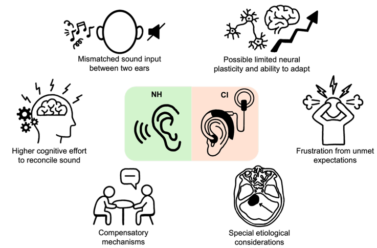
The diagram is a top-down view of the organ of Corti with three rows of outer hair cells (OHC) (represented by gray circles) and a single row of inner hair cells (IHC). The dark gray rectangles represent the supporting cells underneath the hair cell layer. From top to bottom, there is one row of Claudius cells (CC), three rows of Deiters’ cells (DC) and two rows of pillar cells (OPC and IPC). The outer and inner pillar cells are separated by the tunnel of Corti. The top row is a schematic showing how fluorescence (green cells) propagates from the site of damage (OHC marked with red X). The arrow indicates time. The bottom row is the same schematic with a single type II afferent added to show the timing of type II activation relative to the fluorescent wave in the epithelial cells. After noise exposure, the type II afferent calcium-associated fluorescence occurs after the initial epithelial cell wave. Credit: Wood, Megan Beers; Nowak, Nate; Fuchs, Paul Albert/Frontiers in Neurology
In our previous paper in eNeuro in 2021, we showed that an increase in calcium occurred in both epithelial cells and type II afferents after focal damage caused by a laser.
In our new mini-review published in the journal Frontiers in Neurology in February 2024, we focus on cochlear epithelial cells to highlight another tool for studying damage-evoked calcium waves.
There is an increase in fluorescence (a glowing signal) that is visible without calcium-sensitive dyes or genetically encoded calcium indicators.
This signal is thought to result from an increase in the fluorescent molecule NADH. When the calcium wave triggered by cochlear damage enters the mitochondria of cells, it causes an increase in the production of fluorescent NADH from its non-fluorescent form NAD.
The resulting waves of increased fluorescence propagate through tissues similarly (speed and dissipation with distance) to the calcium waves described previously in the cochlea and skin using genetically encoded calcium indicators. Importantly, these waves can be studied in any tissue since no calcium indicators are required.
Of relevance to hyperacusis, prior noise-induced hearing loss leads to the generation of prolonged and repetitive activity in type II neurons and surrounding tissues. This aberrant signaling may be the basis for the sensitivity to everyday sounds seen in hyperacusis. We will continue to explore the activity of epithelial cells and type II cochlear afferent fibers as the cellular basis for pain hyperacusis.
Emerging Research Grants (ERG) scientist Megan Beers Wood, Ph.D., is a postdoctoral research fellow at Johns Hopkins University School of Medicine in the department of otolaryngology. Wood’s 2022 grant was generously funded by Hyperacusis Research, which was renewed for a second year in 2023. She is now a member of Hyperacusis Research’s scientific advisory board. HHF is grateful to Hyperacusis Research for their ongoing support of investigations into noise-induced pain through our ERG program. With their input we have also recently updated our hyperacusis webpage, and HHF friend Julio Flores-Alberca translated it into Spanish.
The Latest Blog Posts


An audiologist shares the science behind Halloween’s eeriest noises.